Human Bone Cross Section Of A Bone - Solved A Human Femur Undergoes Bending And Axial Loading Chegg Com / New users enjoy 60% off.. The large dark spots are passages for blood vessels and nerves. Human bone structure, computer artwork. Cross section of a bone : While it is not as hard as compact bone, spongy bone plays an important role of protecting the marrow where blood cells are produced. Browse 3,292 human bone cross section stock photos and images available or start a new search to explore more stock photos and images.
Each bone in your body is made up of three main types of bone material: Concentric layers of bone cells (osteocytes) and bone matrix surround the central canal. Detailed anatomy of human tooth cross section; Start studying cross section of bone. The central canal, lamellae, canaliculi, and lacunae with osteocytes are apparent.
Cross Section Human Image Photo Free Trial Bigstock from static3.bigstockphoto.com A cross section of a human long bone. Use the illustrations in your textbook as a guide and identify with the scanning objective the following structures. It consists of two layers; Illustration of a cow's hindquarters in cross section. Smooth muscle and endothelium in a muscular artery wall, (magnification x100). Cross section of a bone : Cross‐sectional area is derived from the integral of the bone mass profile across the narrow region. 'delineated as large as the life':
Internal structure of a human long bone, with a magnified cross section of the interior.
Skull cross bone keep fighting never ending hand drawing vector. Red marrow fills the spaces in the spongy bone. Browse 4,275 bone cross section stock photos and images available, or search for human bone cross section to find more great stock photos and pictures. Cross section human cartilage bone microscope view human histological physiology stock photo image by c tonaquatic19 397992404 : Detailed anatomy of human tooth cross section; Smooth muscle and endothelium in a muscular artery wall, (magnification x100). Bone cross section histology : Cross section of a human bone showing bone marrow, spongy bone and blood vessels. Histology slide courtesy of william l. Each epiphysis meets the diaphysis at the metaphysis. Related posts of cross section of human bone diagram human back muscles and bones. Bone is found in the shafts of long bone and consists of various cylindrical units named as haversian system 47. Cross‐sectional area is derived from the integral of the bone mass profile across the narrow region.
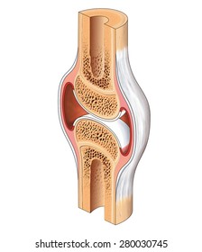
Browse 3,292 human bone cross section stock photos and images available or start a new search to explore more stock photos and images. The large dark spots are passages for blood vessels and nerves. The cortical bone equivalent area of the cross‐section of the region of interest (femoral neck or shaft), with all soft tissue voids (trabecular and cellular spaces) eliminated (cm 2). The central tubular region of the bone, called the diaphysis, flares outward near the end to form the metaphysis, which contains a largely cancellous, or spongy, interior. A cross section of a human long bone.
Bone Human Anatomy from theodora.com Human back muscles and bones 12 photos of the human back muscles and bones human back muscles and bones, bone, human back muscles and bones Bone cross section histology : The cortical bone equivalent area of the cross‐section of the region of interest (femoral neck or shaft), with all soft tissue voids (trabecular and cellular spaces) eliminated (cm 2). Human bone structure, computer artwork. The central tubular region of the bone, called the diaphysis, flares outward near the end to form the metaphysis, which contains a largely cancellous, or spongy, interior. Unlabeled vertebra cross section of human body anatomy infographic diagram including all parts cord of grey and white matter spinal nerve vertebral body foramen and spinous process for medical science. Über 7 millionen englischsprachige bücher. Skull cross bone keep fighting never ending hand drawing vector.
This is known as the periosteum.
Gm1256257085 $ 12.00 istock in stock Concentric layers of bone cells (osteocytes) and bone matrix surround the central canal. The little black spots are osteocytes. Bone test anatomy and physiology 12 photos of the bone test anatomy and physiology anatomy and physiology bone lab test, anatomy and physiology bone markings test, anatomy and physiology bone practical test, anatomy and physiology bone tissue test, anatomy and physiology test on bone tissue, bone, anatomy and. This slide contained a cross section of a very small bone, and you are looking at the entire thickness of the shaft of the bone. Find the perfect human bone cross section stock photo. The central tubular region of the bone, called the diaphysis, flares outward near the end to form the metaphysis, which contains a largely cancellous, or spongy, interior. The central canal, lamellae, canaliculi, and lacunae with osteocytes are apparent. The cortical bone equivalent area of the cross‐section of the region of interest (femoral neck or shaft), with all soft tissue voids (trabecular and cellular spaces) eliminated (cm 2). Skull cross bone keep fighting never ending hand drawing vector. Cross section of a bone / long bone human skeleton fraudbein cross section png 1000x500px watercolor cartoon flower frame heart download free. Items portrayed in this file depicts. Cross section of a human bone showing bone marrow, spongy bone and blood vessels.
The bones serve as levers to the. The large dark spots are passages for blood vessels and nerves. Use the illustrations in your textbook as a guide and identify with the scanning objective the following structures. Start studying cross section of bone. In this short video i use blender 2.8 to show how i created a bone cross section and then use images to control the.
Bone Cross Section High Res Stock Images Shutterstock from image.shutterstock.com Compact bone, spongy bone, and bone marrow. Detailed anatomy of human tooth cross section; This is known as the periosteum. Find the perfect human bone cross section stock illustrations from getty images. Cartilage and bone page 9 from audilab.bmed.mcgill.ca related posts of bone cross section labeled. Fill in the table below. Bone under stereo microscope stereo microscopy is one of the simplest methods to view the surface of a bone. Histology slide courtesy of william l.
Cross section of a human bone showing bone marrow, spongy bone and blood vessels.
Find the perfect human bone cross section stock photo. Detailed anatomy of human tooth cross section; Cross‐sectional area is derived from the integral of the bone mass profile across the narrow region. The large dark spots are passages for blood vessels and nerves. Human bone cross section of a bone. Human bone structure, computer artwork. New users enjoy 60% off. This slide contained a cross section of a very small bone, and you are looking at the entire thickness of the shaft of the bone. Cross section of a bone / long bone human skeleton fraudbein cross section png 1000x500px watercolor cartoon flower frame heart download free. Bone cross section histology : The wider section at each end of the bone is called the epiphysis (plural = epiphyses), which is filled with spongy bone. Concentric layers of bone cells (osteocytes) and bone matrix surround the central canal. Each epiphysis meets the diaphysis at the metaphysis.
Each epiphysis meets the diaphysis at the metaphysis cross section of a bone. Browse 3,292 human bone cross section stock photos and images available or start a new search to explore more stock photos and images.


0 Komentar